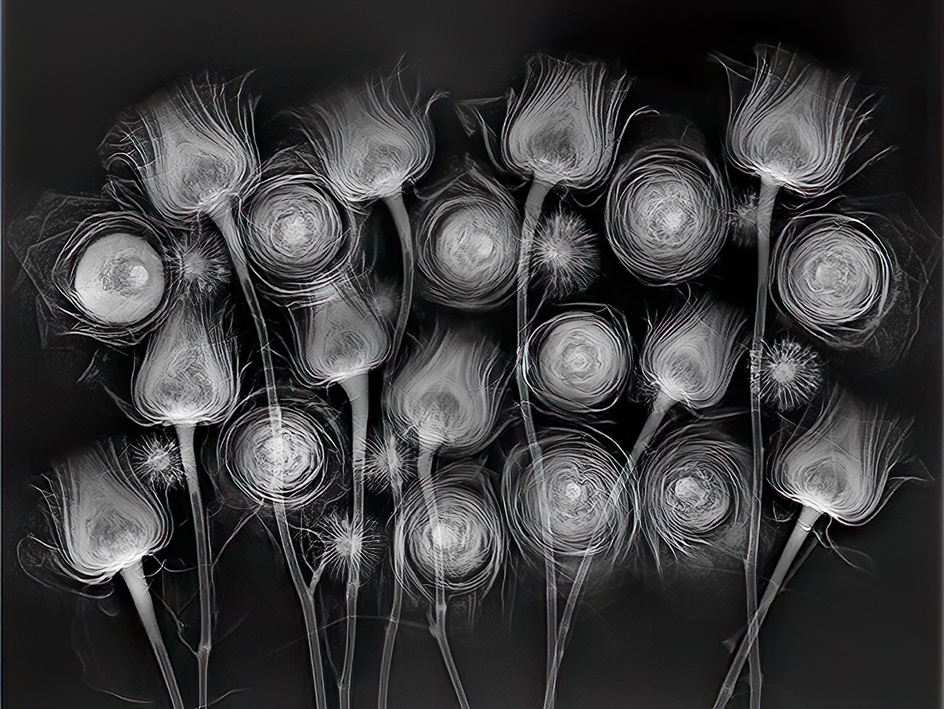
Working with Dr Julian Kopke, I laid out this x-ray composition on a sheet of plexiglass above the sensor. The results you see are actually two x-rays combined, because there is falloff at one of the x-ray, so the second exposure was flipped to create a combined even image. We also used the plexiglass backing in registration to create a light box image of the composition, and I will try later to see what combining the x-ray (interior structure) with the external appearance of the flowers looks like. Check out my FAQ for more information about this kind of imaging.